Segmentation of Retinal Blood Vessels in Fundus Images Using Attention Mechanisms and Deep Supervised Networks
DOI:
https://doi.org/10.21512/commit.v20i1.13174Keywords:
Retinal Blood Vessels, Diabetic Retinopathy, Deep Supervision, Semantic Segmentation, Attention MechanismAbstract
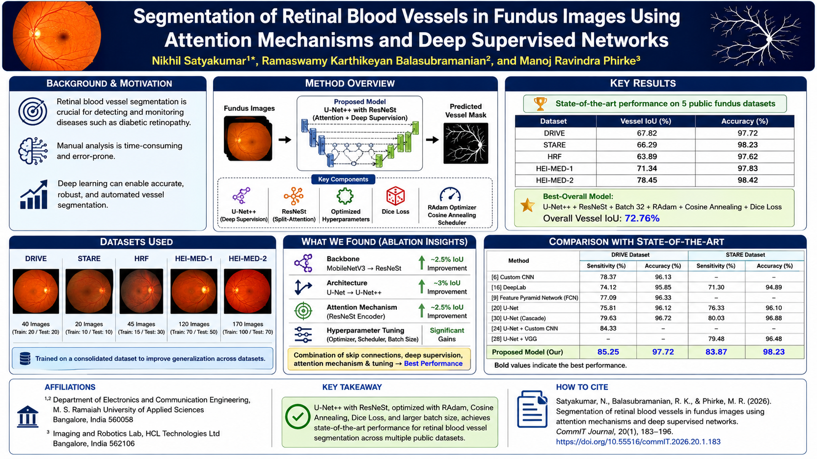
Retinal blood vessel segmentation is crucial for detecting and monitoring retinal disorders such as diabetic retinopathy, age-related macular degeneration and glaucoma. Automating the segmentation of blood vessels leads to a reduction in the time and cost of manual segmentation, enables large-scale clinical studies, improves accuracy, ensures consistency, allows for realtime analysis, and facilitates early disease detection. The research examines the performance of 7 semantic segmentation architectures, each combined with 10 pretrained backbones, on 5 publicly available fundus image datasets. Models are trained on a NVIDIA GeForce GTX 1080 Graphics Processing Unit (GPU), and key hyperparameters, such as batch size, optimizers, and learning rate schedulers, are systematically optimized. Intersection over Union (IoU), accuracy, sensitivity, and computational time are used as key performance indicators. Approximately 97 experiments are conducted to achieve state-of-the-art accuracies of 97.72%, 98.23%, 97.62%, 97.83%, and 98.42%, along with IoU scores of 67.82%, 66.29%, 63.89%, 71.34%, and 78.45% on the DRIVE, STARE, HRF, HEI-MED-1, and HEI-MED-2 datasets, respectively. The best performance is achieved using the U-Net++ architecture with ResNeSt backbone, RAdam optimizer, and Cosine Annealing scheduler. This combination leverages deep supervision, attention mechanisms, and bottleneck architectures to enhance multiscale feature learning, localization, robustness to image variability, and model generalization. Although the models demonstrate strong performance, challenges remain in addressing dataset imbalance and ensuring generalization to unseen patient populations.
![]()
References
[1] A. E. Fayed, M. J. Menten, L. Kreitner, J. C. Paetzold, D. Rueckert, S. M. Bassily, R. R. Fikry, A. M. Hagag, and S. Sivaprasad, “Retinal vasculature of different diameters and plexuses exhibit distinct vulnerability in varying severity of diabetic retinopathy,” Eye, vol. 38, no. 9, pp. 1762–1769, 2024.
[2] Z. Liu, M. S. Sunar, T. S. Tan, and W. H. W. Hitam, “Deep learning for retinal vessel segmentation: A systematic review of techniques and applications,” Medical & Biological Engineering & Computing, vol. 63, no. 8, pp. 2191–2208, 2025.
[3] Y. Gao, Y. Jiang, Y. Peng, F. Yuan, X. Zhang, and J. Wang, “Medical image segmentation: A comprehensive review of deep learning-based methods,” Tomography, vol. 11, no. 5, pp. 1–45, 2025.
[4] L. Ming and L. Qi, “DMSU-Net++: A dual multiscale retinal vessel segmentation method based on improved U-Net++,” PLOS ONE, vol. 20, no. 7, pp. 1–13, 2025.
[5] M. Zhang, F. Yu, J. Zhao, L. Zhang, and Q. Li, “BEFD: Boundary enhancement and feature denoising for vessel segmentation,” in International Conference on Medical Image Computing and Computer-Assisted Intervention. Lima, Peru: Springer, Oct. 4–8, 2020, pp. 775–785.
[6] L. Yu, Z. Qin, T. Zhuang, Y. Ding, Z. Qin, and K. K. R. Choo, “A framework for hierarchical division of retinal vascular networks,” Neurocomputing, vol. 392, pp. 221–232, 2020.
[7] V. Cherukuri, V. K. Bg, R. Bala, and V. Monga, “Deep retinal image segmentation with regularization under geometric priors,” IEEE Transactions on Image Processing, vol. 29, pp. 2552–2567, 2019.
[8] J. Ding, Z. Zhang, J. Tang, and F. Guo, “A multichannel deep neural network for retina vessel segmentation via a fusion mechanism,” Frontiers in Bioengineering and Biotechnology, vol. 9, pp. 1–14, 2021.
[9] Z. Feng, J. Yang, and L. Yao, “Patch-based fully convolutional neural network with skip connections for retinal blood vessel segmentation,” in 2017 IEEE International Conference on Image Processing (ICIP). Beijing, China: IEEE, Sep. 17–20, 2017, pp. 1742–1746.
[10] Y. Jiang, F. Wang, J. Gao, and W. Liu, “Efficient BFCN for automatic retinal vessel segmentation,” Journal of Ophthalmology, vol. 2020, no. 1, pp. 1–14, 2020.
[11] Y. Zhong, T. Chen, D. Zhong, and X. Liu, “Wavelet-guided network with fine-grained feature extraction for vessel segmentation.” The Visual Computer, vol. 41, no. 6, pp. 4377–4392, 2025.
[12] Y. Liu, J. Shen, L. Yang, H. Yu, and G. Bian, “Wave-Net: A lightweight deep network for retinal vessel segmentation from fundus images,” Computers in Biology and Medicine, vol. 152, 2023.
[13] J. Ryu, M. U. Rehman, I. F. Nizami, and K. T. Chong, “SegR-Net: A deep learning framework with multi-scale feature fusion for robust retinal vessel segmentation,” Computers in Biology and Medicine, vol. 163, 2023.
[14] N. Mukkapati and M. S. Anbarasi, “Brain tumor classification based on enhanced CNN model,” Revue d’Intelligence Artificielle, vol. 36, no. 1, pp. 125–130, 2022.
[15] Y. Bai, J. Li, L. Shi, Q. Jiang, B. Yan, and Z. Wang, “DME-DeepLabV3+: A lightweight model for diabetic macular edema extraction based on DeepLabV3+ architecture,” Frontiers in Medicine, vol. 10, pp. 1–11, 2023.
[16] H. Fu, Y. Xu, S. Lin, D. W. Kee Wong, and J. Liu, “Deepvessel: Retinal vessel segmentation via deep learning and conditional random field,” in International Conference on Medical Image Computing and Computer-Assisted Intervention. Athens, Greece: Springer, Oct. 17–21, 2016, pp. 132–139.
[17] H. J. Kim, H. Eesaar, and K. T. Chong, “Transformer-enhanced retinal vessel segmentation for diabetic retinopathy detection using attention mechanisms and multi-scale fusion,” Applied Sciences, vol. 14, no. 22, pp. 1–19, 2024.
[18] S. Kushwaha, J. Boga, B. S. S. Rao, S. N. Taqui, R. G. Vidhya, and J. Surendiran, “Machine learning method for the diagnosis of retinal diseases using convolutional neural network,” in 2023 International Conference on Data Science, Agents & Artificial Intelligence (ICDSAAI). Chennai, India: IEEE, Dec. 21–23, 2023, pp. 1–6.
[19] Z. Li, M. Jia, X. Yang, and M. Xu, “Blood vessel segmentation of retinal image based on dense-UNet network,” Micromachines, vol. 12, no. 12, pp. 1–11, 2021.
[20] Y. Wu, Y. Xia, Y. Song, D. Zhang, D. Liu, C. Zhang, and W. Cai, “Vessel-Net: Retinal vessel segmentation under multi-path supervision,” in International Conference on Medical Image Computing and Computer-Assisted Intervention. Shenzhen, China: Springer, Oct. 13–17, 2019, pp. 264–272.
[21] Z. Huang, Y. Fang, H. Huang, X. Xu, J. Wang, and X. Lai, “Automatic retinal vessel segmentation based on an improved U-Net approach,” Scientific Programming, vol. 2021, no. 1, pp. 1–15, 2021.
[22] H. Hu and Z. Liu, “Retinal vessel segmentation based on recurrent convolutional skip connection U-Net,” in 2021 4th International Conference on Intelligent Autonomous Systems (ICoIAS). Wuhan, China: IEEE, May 14–16, 2021, pp. 65–71.
[23] S. Mishra, D. Z. Chen, and X. S. Hu, “A dataaware deep supervised method for retinal vessel segmentation,” in 2020 IEEE 17th International Symposium on Biomedical Imaging (ISBI). Iowa City, IA, USA: IEEE, April 3–7, 2020, pp. 1254–1257.
[24] H. Zhao, H. Li, and L. Cheng, “Improving retinal vessel segmentation with joint local loss by matting,” Pattern Recognition, vol. 98, 2020.
[25] X. Du, J. Wang, and W. Sun, “Densely connected U-Net retinal vessel segmentation algorithm based on multi-scale feature convolution extraction,” Medical Physics, vol. 48, no. 7, pp. 3827–3841, 2021.
[26] C. Liu, P. Gu, and Z. Xiao, “Multiscale U-Net with spatial positional attention for retinal vessel segmentation,” Journal of Healthcare Engineering, vol. 2022, no. 1, pp. 1–10, 2022.
[27] X. Yang, L. Liu, and T. Li, “MR-UNet: An UNet model using multi-scale and residual convolutions for retinal vessel segmentation,” International Journal of Imaging Systems and Technology, vol. 32, no. 5, pp. 1588–1603, 2022.
[28] S. Feng, Z. Zhuo, D. Pan, and Q. Tian, “Cc-Net: A cross-connected convolutional network for segmenting retinal vessels using multi-scale features,” Neurocomputing, vol. 392, pp. 268–276, 2020.
[29] X. Li, J. Ding, J. Tang, and F. Guo, “Res2Unet: A multi-scale channel attention network for retinal vessel segmentation,” Neural Computing and Applications, vol. 34, no. 14, pp. 12 001–12 015, 2022.
[30] Y. Wu, Y. Xia, Y. Song, Y. Zhang, and W. Cai, “NFN+: A novel network followed network for retinal vessel segmentation,” Neural Networks, vol. 126, pp. 153–162, 2020.
[31] Y. Wu, A. Liu, L. Chen, D. Zhao, H. Zhou, and Q. Zheng, “Multi-scale attention net for retina blood vessel segmentation,” in Proceedings of the 2020 4th International Conference on Computer Science and Artificial Intelligence. Zhuhai. China: Association for Computing Machinery, Dec. 11–13, 2020, pp. 86–90.
[32] G. Wang, Y. Huang, K. Ma, Z. Duan, Z. Luo, P. Xiao, and J. Yuan, “Automatic vessel crossing and bifurcation detection based on multi-attention network vessel segmentation and directed graph search,” Computers in Biology and Medicine, vol. 155, pp. 1–11, 2023.
[33] C. Kromm and K. Rohr, “Inception capsule network for retinal blood vessel segmentation and centerline extraction,” in 2020 IEEE 17th International Symposium on Biomedical Imaging (ISBI). IEEE, 2020, pp. 1223–1226.
[34] K. B. Khan, M. S. Siddique, M. Ahmad, and M. Mazzara, “A hybrid unsupervised approach for retinal vessel segmentation,” BioMed Research International, vol. 2020, no. 1, pp. 1–20, 2020.
[35] L. C. Chen, G. Papandreou, I. Kokkinos, K. Murphy, and A. L. Yuille, “DeepLab: Semantic image segmentation with deep convolutional nets, atrous convolution, and fully connected CRFs,” IEEE transactions on pattern analysis and machine intelligence, vol. 40, no. 4, pp. 834–848, 2017.
[36] Q. Zhao, J. Cao, J. Ge, Q. Zhu, X. Chen, and W. Liu, “Multi-UNet: An effective Multi-U convolutional networks for semantic segmentation,” Knowledge-Based Systems, vol. 309, 2025.
[37] L. C. Chen, Y. Zhu, G. Papandreou, F. Schroff, and H. Adam, “Encoder-decoder with atrous separable convolution for semantic segmentation,” in Computer Vision – ECCV 2018, Munich, Germany, Sep. 8–14, 2018.
[38] Z. Zhou, M. M. Rahman Siddiquee, N. Tajbakhsh, and J. Liang, “UNet++: A nested U-Net architecture for medical image segmentation,” in International Workshop on Deep Learning in Medical Image Analysis. Granada, Spain: Springer, Sep. 20, 2018, pp. 3–11.
[39] A. Chaurasia and E. Culurciello, “LinkNet: Exploiting encoder representations for efficient semantic segmentation,” in 2017 IEEE Visual Communications and Image Processing (VCIP). St. Petersburg, FL, USA: IEEE, Dec. 10–13, 2017, pp. 1–4.
[40] T. Fan, G. Wang, Y. Li, and H. Wang, “Ma-Net: A multi-scale attention network for liver and tumor segmentation,” IEEE Access, vol. 8, pp. 179 656–179 665, 2020.
[41] M. Hasal, M. Pecha, J. Nowakov´a, D. Hern´andez-Sosa, V. Sn´aˇsel, and J. Timkoviˇc, “Retinal vessel segmentation by U-Net with VGG-16 backbone on patched images with smooth blending,” in International Conference on Intelligent Networking and Collaborative Systems. Springer, 2023, pp. 465–474.
[42] Y. Chen, J. Li, H. Xiao, X. Jin, S. Yan, and J. Feng, “Dual path networks,” in NIPS’17: Proceedings of the 31st International Conference on Neural Information Processing Systems. Long Beach California, USA: Curran Associates Inc., Dec. 4–9, 2017, pp. 4470–4478.
[43] C. Szegedy, S. Ioffe, V. Vanhoucke, and A. Alemi, “Inception-v4, Inception-ResNet and the impact of residual connections on learning,” in AAAI’17: Proceedings of the Thirty-First AAAI Conference on Artificial Intelligence. San Francisco, USA: Association for the Advancement of ArtificialIntelligence, Feb. 4–9, 2017, pp. 4278–4284.
[44] X. Jin, Y. Xie, X. S. Wei, B. R. Zhao, Z. M. Chen, and X. Tan, “Delving deep into spatial pooling for squeeze-and-excitation networks,” Pattern Recognition, vol. 121, 2022.
[45] S. Qian, C. Ning, and Y. Hu, “MobileNetV3 for image classification,” in 2021 IEEE 2nd International Conference on Big Data, Artificial Intelligence and Internet of Things Engineering (ICBAIE). Nanchang, China: IEEE, March 26–28, 2021, pp. 490–497.
[46] M. Lin, H. Chen, X. Sun, Q. Qian, H. Li, and R. Jin, “Neural architecture design for GPUefficient networks,” 2020. [Online]. Available: https://arxiv.org/abs/2006.14090
[47] H. Zhang, C. Wu, Z. Zhang, Y. Zhu, H. Lin, Z. Zhang, Y. Sun, T. He, J. Mueller, R. Manmatha, M. Li, and A. Smola, “ResNeSt: Splitattention networks,” in Proceedings of the IEEE/CVF Conference on Computer Vision and Pattern Recognition, New Orleans, Louisiana, 2022, pp. 2736–2746.
[48] I. Radosavovic, R. P. Kosaraju, R. Girshick, K. He, and P. Doll´ar, “Designing network design spaces,” in Proceedings of the IEEE/CVF Conference on Computer Vision and Pattern Recognition, 2020, pp. 10 428–10 436.
[49] D. P. Kingma and J. Ba, “Adam: A method for stochastic optimization,” 2014. [Online]. Available: https://arxiv.org/abs/1412.6980
[50] A. Graves, “Generating sequences with recurrent neural networks,” 2013. [Online]. Available: https://arxiv.org/abs/1308.0850
[51] T. Dozat, “Incorporating Nesterov momentum into Adam,” 2016. [Online]. Available: https://openreview.net/forum?id=OM0jvwB8jIp57ZJjtNEZ
[52] I. Loshchilov and F. Hutter, “Decoupled weight decay regularization,” 2017. [Online]. Available: https://arxiv.org/abs/1711.05101
[53] L. Liu, H. Jiang, P. He, W. Chen, X. Liu, J. Gao, and J. Han, “On the variance of the adaptive learning rate and beyond,” 2019. [Online]. Available: https://arxiv.org/abs/1908.03265
[54] T. P. T. Armand, S. Bhattacharjee, H.-K. Choi, and H. C. Kim, “Transformers effectiveness in medical image segmentation: A comparative analysis of UNet-based architectures,” in 2024 International Conference on Artificial Intelligence in Information and Communication (ICAIIC). Osaka, Japan: IEEE, Feb. 19–22, 2024, pp. 238–242.
Downloads
Published
How to Cite
Issue
Section
License
Copyright (c) 2026 Nikhil Satyakumar, Ramaswamy Karthikeyan Balasubramanian, Manoj Ravindra Phirke

This work is licensed under a Creative Commons Attribution-ShareAlike 4.0 International License.
Authors who publish with this journal agree to the following terms:
a. Authors retain copyright and grant the journal right of first publication with the work simultaneously licensed under a Creative Commons Attribution License - Share Alike that allows others to share the work with an acknowledgment of the work's authorship and initial publication in this journal.
b. Authors are able to enter into separate, additional contractual arrangements for the non-exclusive distribution of the journal's published version of the work (e.g., post it to an institutional repository or publish it in a book), with an acknowledgment of its initial publication in this journal.
c. Authors are permitted and encouraged to post their work online (e.g., in institutional repositories or on their website) prior to and during the submission process, as it can lead to productive exchanges, as well as earlier and greater citation of published work.
Â
USER RIGHTS
All articles published Open Access will be immediately and permanently free for everyone to read and download. We are continuously working with our author communities to select the best choice of license options, currently being defined for this journal as follows: Creative Commons Attribution-Share Alike (CC BY-SA)